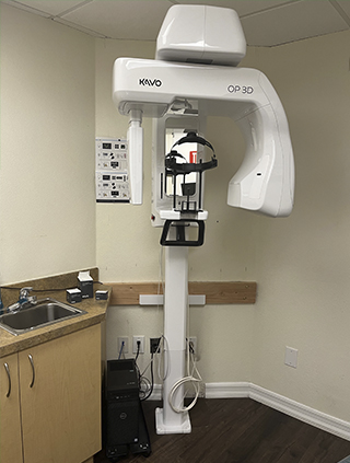
In the ever-evolving landscape of modern dentistry, technology continues to play a pivotal role in enhancing diagnostic accuracy, treatment planning, and patient care. One such revolutionary advancement is Cone Beam Computed Tomography (CBCT), a sophisticated imaging technique that has transformed the way dentists perceive and address oral health concerns.
What is CBCT?
Cone Beam Computed Tomography, often abbreviated as CBCT, is a specialized medical imaging technique that provides three-dimensional views of the oral and maxillofacial regions. Unlike traditional two-dimensional X-rays, CBCT captures high-resolution, cross-sectional images of a patient's teeth, jawbones, and surrounding structures. This technology utilizes a cone-shaped X-ray beam to generate a comprehensive image, allowing dental professionals to visualize intricate details that might go unnoticed with conventional radiographs.
Applications in Dentistry
CBCT imaging has become an indispensable tool across various dental specialties, offering precise information for diagnosis, treatment planning, and monitoring of numerous oral conditions. This includes applications in implant dentistry, endodontics, oral and maxillofacial surgery, orthodontics, and TMJ (Temporomandibular Joint) disorders.
In implant dentistry, CBCT scans prove invaluable for assessing bone density, volume, and anatomical suitability for dental implant placement. Dentists can plan implant procedures with utmost precision, minimizing risks and ensuring long-term success.
In endodontics, CBCT helps identify complex root canal anatomy, locate hidden canals, and diagnose issues that may not be apparent in regular X-rays, leading to more effective and successful root canal therapies.
Oral and maxillofacial surgeons use CBCT to plan complex surgeries such as impacted wisdom tooth extractions, orthognathic surgeries (jaw corrections), and the removal of cysts or tumors. This aids in minimizing surgical trauma and improving post-operative outcomes.
Orthodontists utilize CBCT to assess the position of teeth, root angulations, and jaw relationships in three dimensions. This information is crucial for designing customized orthodontic treatment plans, including braces or clear aligners.
In addressing TMJ disorders, CBCT provides detailed images of the TMJ, aiding in diagnosis and management. This helps tailor treatment approaches to individual patient needs effectively.
The adoption of CBCT technology in dentistry offers several distinct advantages. These include enhanced diagnostic accuracy, reduced radiation exposure compared to traditional CT scans, non-invasiveness, time efficiency, and improved treatment outcomes.
In conclusion, Cone Beam Computed Tomography (CBCT) has revolutionized dentistry by offering precise 3D images, improving diagnoses, and enabling better treatment planning. This technology has not only improved patient care but has also expanded the horizons of what is possible in the field of dentistry. As CBCT technology continues to evolve, we can expect even more remarkable advancements in the years to come, further enhancing the quality of dental care worldwide.
Please visit Safe Dental Care in Oxnard, CA today for a consultation.
